Plasma and UV-light surface modification of Titanium Surfaces
Effects of Surface Modification on Adsorption Behavior of Cell and Protein on Titanium Surface by Using Quartz Crystal Microbalance System
Abstract
The effect of plasma and UV-light treatment of titanium surfaces on the adsorption behaviour of cells and proteins was investigated. With SEM and SPM images show that the microstructure remains unaffected. Meanwhile, the surface composition changes according to XPS measurements from a high amount of carbonous material to a more blank titanium surface. Along this the surface wettability is strongly increased. With a quartz crystal microbalance the adsorption behaviour of cells was investigated, indicating an intermediate effect on the biocompatibility for the UV-treated samples, and a strong effect for the plasma-treated samples.
Authors: Matsumoto, T.; Tashiro, Y.; Komasa, S.; Miyake, A.; Komasa, Y. & Okazaki, J.
Publication: Effects of Surface Modification on Adsorption Behavior of Cell and Protein on Titanium Surface by Using Quartz Crystal Microbalance System, Materials, 2021, 14.
In a recent publication Matsumoto et al. compared the effect of ultraviolet light and atmospheric plasma on the adsorption behavior of cells and proteins on a titanium surface. Titanium and its alloys is the most commonly used material for dental implants. The osseointegration is critical for the successful clinical outcome, which can take several months after implantation. Especially the primary stability is based on the speed of adsorption. In this investigation the deposition of albumin, fibronectin, and rat bone marrow (RBM) cells on titanium disks was monitored with a quartz crystal microbalance. The titanium plates were prepared in three different ways: As a reference an untreated plate was used. The other fraction was treated with UV-light (HL-2000 HybriLinker; Funakoshi, Tokyo, Japan) for 15 min. The third fraction was treated with cold atmospheric plasma (piezobrush® PZ2, relyon plasma, Germany) for 30 s at 10 mm distance. These samples were characterized with SEM, XPS and contact angle measurements. Furthermore, the samples were exposed to the proteins while the adsorped mass was monitored with a QCM (Affinix QNµ; Initium Co., Ltd., Tokyo, Japan).
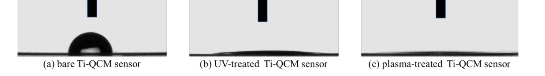
The SEM and SPM images show that there is no significant change in the surface microstructure by the pre-treating with UV-radiation and cold plasma. Especially the surface roughness is for all three samples very comparable. This indicates that no structural changes are introduced to the surface. The XPS however, shows a shift in the elemental composition. Especially the oxygen contribution is increased by the UV- and plasma treatment compared to the bare probe. Meanwhile the carbon content is lowered for the treated samples. This trend is very pronounced for the plasma treated samples. A similar tendency can be found for the contact angle measurement. The water contact angle of the untreated sample indicates with 90.6 ° an hydrophobic behaviour. It could be lowered with UV to 7.2 ° and with plasma to complete wetting (0 °). The hydrophilicity of the treated samples was attributed to the removal of carbon compounds via the pre-treatments.
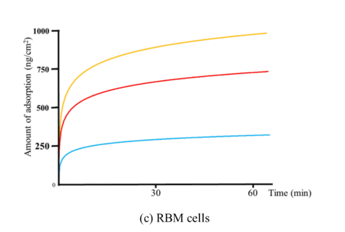
The measurements with the QCM show for all investigated cell types the fastest adsorption for the plasma treated samples, followed by the UV-treated and the untreated samples. The same is true for the total adsorbed weight after 60 minutes. A similar trend can be seen for the mineralization, where the calcium deposition on RBM cells seeded on the untreated, UV-treated and plasma-treated titanium disks was determined after 21 and 28 days. As an other indication for the osseointegration the ROS accumulation was monitored. The same trend as for the other measurements could be found here.
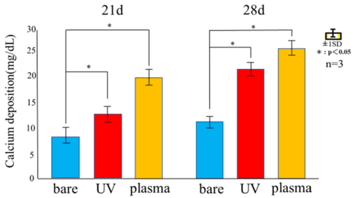
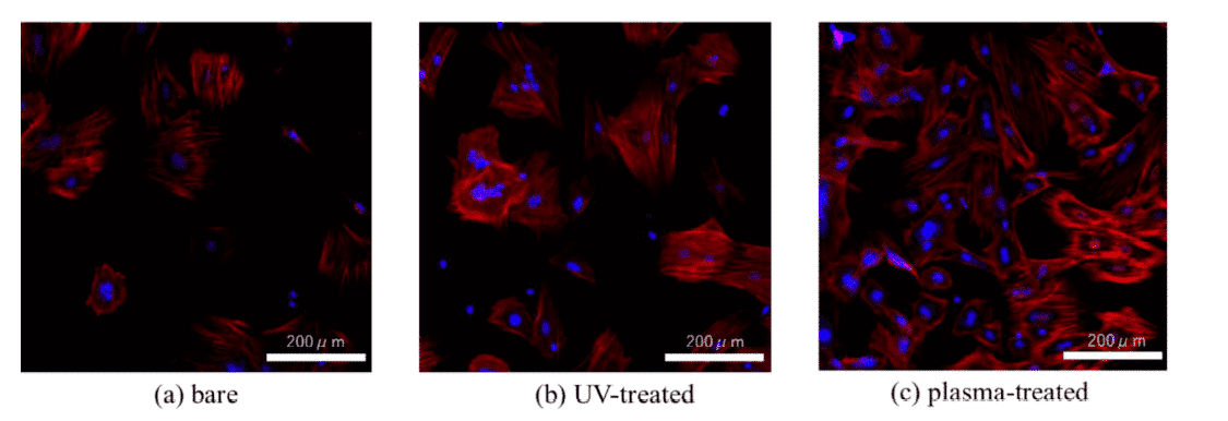
The effectiveness of UV-radiation and cold atmospheric plasma for hydrophilizing implant surfaces was evaluated by simulating the initial hard-tissue-formation behavior of the tissue surrounding the implant material. The surface morphology remains unchanged upon treatment, however, the wettability is highly increased by both methods. The plasma-treated titanium surface showed the lowest contact angle, which enabled the highest adsorption of albumin, fibronectin, and RBM cells. While the performance of the UV-treated and plasma-treated samples were generally similar, the plasma-treated samples showed the highest amounts of F-actin, filopodia, and lamellipodia, the highest ALP activity, the highest amount of calciumprecipitation, and the lowest ROS value of all sample types.
Read the whole report here.